Ultrasound shear wave elastography functions as a critical, non-invasive instrument for quantifying tissue mechanics. It contributes to foot biomechanical research by measuring the speed of shear wave propagation to generate 2D maps of tissue stiffness, effectively identifying localized hardening caused by repetitive loading before visible injury occurs.
Core Takeaway Ultrasound shear wave elastography fills the diagnostic gap between healthy tissue and macroscopic injury. By detecting early increases in shear wave velocity—a proxy for stiffness—it allows researchers to identify internal biomechanical changes and assess the degree of tissue damage prior to structural failure.
The Mechanics of Stiffness Quantification
Measuring Wave Propagation
The fundamental metric of this technology is the propagation speed of shear waves.
By analyzing how quickly these waves travel through soft tissue, researchers can derive concrete data regarding tissue density and tension.
Creating the 2D Stiffness Map
Rather than providing a single data point, this method generates a 2D distribution map.
This allows for the visualization of stiffness gradients across the plantar soft tissue, highlighting specific areas of concern rather than generalized averages.
Correlating Speed with Hardness
There is a direct relationship between wave velocity and tissue state.
An increase in shear wave velocity indicates tissue hardening. This quantitative link provides an objective baseline for assessing the mechanical health of the foot.
Early Detection and Injury Prevention
Identifying Repetitive Loading Effects
In foot biomechanics, tissues often degrade or harden due to cumulative stress.
Elastography effectively detects the localized tissue hardening that results from this repetitive loading, a subtle change that traditional imaging might miss.
Pre-Macroscopic Diagnosis
The most significant contribution of this technology is its predictive capability.
It identifies early biomechanical changes in internal tissues before macroscopic injury occurs, shifting the focus from reactive treatment to proactive prevention.
Understanding the Scope of Assessment
Focus on Stiffness vs. Anatomy
While traditional ultrasound looks at structure, shear wave elastography focuses specifically on quantitative stiffness.
It is a functional assessment tool designed to grade the degree of tissue damage based on elasticity, rather than simply visualizing anatomical disruptions.
Sensitivity to Localized Changes
The effectiveness of this tool relies on its ability to detect localized anomalies.
Researchers must interpret the 2D maps carefully to isolate specific hardened regions from the surrounding healthy tissue to accurately assess the pathology.
Applying These Insights to Research
To maximize the value of ultrasound shear wave elastography in your work, consider your specific analytical goals:
- If your primary focus is Injury Prevention: Monitor changes in shear wave velocity to detect hardening from repetitive loading before physical symptoms appear.
- If your primary focus is Damage Assessment: Use the 2D stiffness distribution maps to quantitatively grade the severity of tissue damage in injured patients.
This technology transforms subjective palpation into objective data, providing a definitive metric for soft tissue health.
Summary Table:
| Feature | Description | Biomechanical Benefit |
|---|---|---|
| Wave Propagation | Measures speed of shear waves through tissue | Quantifies tissue density and mechanical tension |
| 2D Stiffness Map | Visualizes stiffness gradients across the foot | Pinpoints localized hardening from repetitive loading |
| Quantitative Metric | Correlates wave velocity with tissue hardness | Provides objective data before macroscopic injury occurs |
| Early Detection | Identifies subtle changes in internal tissues | Shifts focus from reactive treatment to proactive prevention |
Elevate Your Footwear Performance with 3515
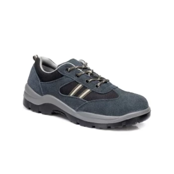
As a large-scale manufacturer serving global distributors and brand owners, 3515 leverages advanced biomechanical insights to produce superior footwear. Our comprehensive production capabilities cover all categories—from our flagship Safety Shoes series and tactical boots to outdoor, training, and dress shoes.
By understanding the science of tissue mechanics, we design footwear that minimizes repetitive loading stress and maximizes foot health. Partner with us to bring scientifically-backed footwear to your market.
Contact our expert team today to discuss your bulk requirements!
References
- Panagiotis Chatzistergos, Nachiappan Chockalingam. An in vivo model for overloading-induced soft tissue injury. DOI: 10.1038/s41598-022-10011-7
This article is also based on technical information from 3515 Knowledge Base .
Related Products
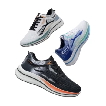
- Lightweight Breathable Sneakers with Wet-Traction Grip for Wholesale & Private Label
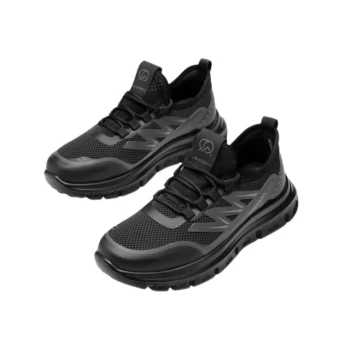
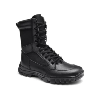
- Wholesale Durable Safety Boots Manufacturer Customizable Steel Toe Work Boots
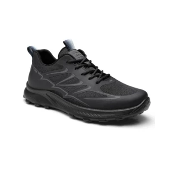
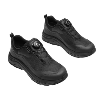
- Lightweight Breathable Training Shoes for Wholesale & Custom OEM Manufacturing
- Wholesale Durable & Breathable Training Shoes for Custom Brands
- Wholesale Breathable & Cushioned Training Shoes Custom Factory Production
People Also Ask
- What are the benefits of wearing lightweight shoes? Reduce Fatigue & Boost Comfort for Work & Play
- Why is breathability important in shoes for all-day wear? Prevent Sweat, Blisters, and Fatigue
- How do lightweight shoes provide support? The Science of Efficient Cushioning & Stability
- Why are breathable sneakers necessary for tropical commuting? Stay Cool and Dry in High Humidity
- What are the style advantages of lightweight shoes? Sleek, Versatile & Modern Comfort