Marking patches serve as the critical link between physical anatomy and digital reconstruction in 3D foot scanning. By physically attaching these markers to anatomical landmarks identified through manual palpation, you provide the scanning system with explicit spatial references. This ensures the software can accurately map surface coordinates to the underlying deep skeletal structures that are otherwise invisible to the camera.
Core Takeaway 3D scanners capture surface geometry, but biomechanics rely on skeletal alignment. Marking patches bridge this gap by translating palpable bone landmarks—like the navicular bone—into digital data points, ensuring that calculated parameters reflect anatomical reality rather than just surface shape.
Bridging the Gap Between Surface and Skeleton
Connecting Palpation to Digital Data
A 3D scanner is an optical tool; it can only record the skin's surface. However, true biomechanical analysis requires data on the skeletal structure beneath.
To solve this, an operator must first perform palpation to locate specific bony landmarks, such as the navicular bone point or the highest point of the talus head.
Once these points are found physically, marking patches are applied to "flag" these locations. This converts a tactile, human assessment into a visual cue that the digital scanner can record.
Identifying Deep Structures
Without markers, a scanner treats the foot as a continuous, uniform surface. It cannot distinguish where a bone protrudes versus where soft tissue swells.
Marking patches provide explicit spatial references. They tell the scanning program exactly which surface positions correspond to deep skeletal structures.
This allows the software to anchor its digital reconstruction to the actual anatomy of the patient, rather than relying on algorithmic estimation of where bones "should" be.
Improving Biomechanical Reliability
Enhancing Complex Geometry Analysis
Foot geometries are complex and vary significantly between individuals. Relying solely on surface shape can lead to misinterpretation of the foot's mechanics.
Markers ensure the scanning program correctly identifies critical zones. This is particularly vital when analyzing feet with deformities or unusual structures where standard anatomical assumptions might fail.
Calculating Critical Parameters
The ultimate goal of using these markers is to improve the reliability of the output data.
The primary reference highlights that markers significantly improve the calculation of critical biomechanical parameters. Specifically, they ensure accuracy in measuring:
- Arch height: The true vertical distance from the skeletal anchor.
- Transverse arch width: Accurate spacing between bony landmarks.
- Midfoot rotation torque: Precise rotational measurements derived from fixed skeletal points.
Understanding the Trade-offs
Dependence on Manual Skill
While marking patches improve digital accuracy, they introduce a dependency on manual proficiency.
The reliability of the 3D scan becomes directly tied to the accuracy of the initial palpation. If the operator misidentifies the navicular bone or talus head physically, the "precise" digital marker will lock the system into an incorrect reference point.
The "Garbage In, Garbage Out" Risk
The scanner trusts the marker implicitly. It assumes the patch represents the true skeletal structure.
Therefore, the system provides no safeguard against human error during the pre-scan setup. The technology enhances the reliability of the calculation, but it cannot validate the location of the marker itself.
Making the Right Choice for Your Goal
To determine when and how to utilize marking patches effectively, consider your specific analytical needs:
- If your primary focus is deep biomechanical analysis: You must use markers on the navicular bone and talus head to obtain reliable data on torque and arch mechanics.
- If your primary focus is surface topology: You may not need markers, but understand that your data will lack specific skeletal referencing.
Use marking patches to force your digital tools to respect the physical reality of the patient's skeletal structure.
Summary Table:
| Feature | Role of Marking Patches | Benefit to 3D Scanning |
|---|---|---|
| Data Bridge | Links manual palpation to digital data | Translates physical bone landmarks into digital points |
| Structural ID | Identifies deep skeletal structures | Distinguishes bone protrusions from soft tissue swelling |
| Parameter Accuracy | Anchors calculations for arches & torque | Ensures reliable measurement of arch height and rotation |
| Geometry Mapping | Provides explicit spatial references | Corrects for unique foot deformities or complex shapes |
Elevate Your Footwear Production with 3515’s Precision Engineering
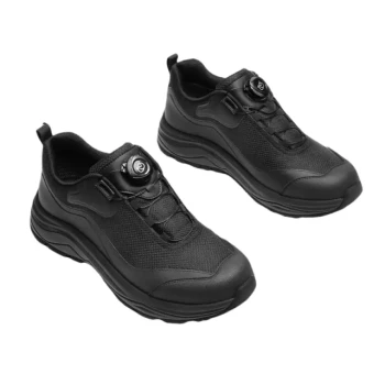
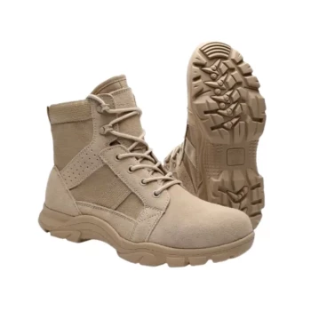
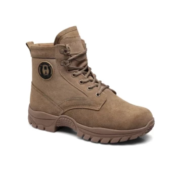
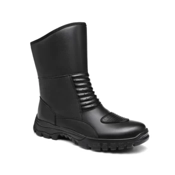
As a large-scale manufacturer serving distributors and brand owners, 3515 offers comprehensive production capabilities for all footwear types. Our flagship Safety Shoes series is built on the same principles of anatomical precision and biomechanical reliability discussed above, ensuring every pair provides optimal support and protection.
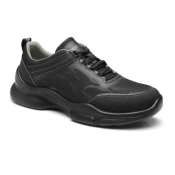
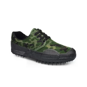
Our extensive portfolio covers everything from work and tactical boots to outdoor shoes, training shoes, sneakers, and Dress & Formal shoes, designed to meet your diverse bulk requirements with uncompromising quality. Partner with a manufacturer that understands the science of the perfect fit.
Contact 3515 Today to Discuss Your Bulk Footwear Needs
References
- Tomoko Yamashita, Shingo Ata. Evaluation of Hallux Valgus Using Rotational Moment of Midfoot Measured by a Three-dimensional Foot Scanner: a Cross-sectional Observational Study. DOI: 10.14326/abe.12.154
This article is also based on technical information from 3515 Knowledge Base .
Related Products
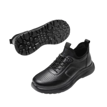
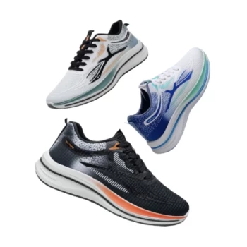
- Lightweight Breathable Sneakers with Wet-Traction Grip for Wholesale & Private Label
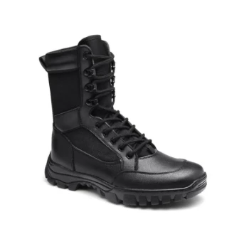
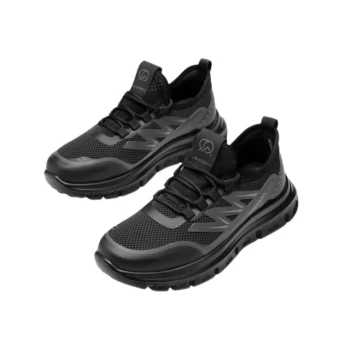
- Wholesale Durable Safety Boots Manufacturer Customizable Steel Toe Work Boots
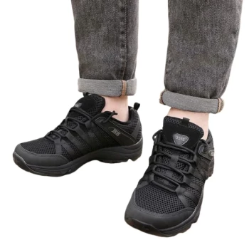
- Lightweight Breathable Training Shoes for Wholesale & Custom OEM Manufacturing
- Wholesale Durable & Breathable Training Shoes for Custom Brands
- Wholesale Comfortable Business Casual Shoes Custom Manufacturing
People Also Ask
- How do lightweight shoes provide support? The Science of Efficient Cushioning & Stability
- Why are lightweight training shoes often preferred over professional hiking boots in urban walking? Maximize City Comfort
- Why are breathable sneakers necessary for tropical commuting? Stay Cool and Dry in High Humidity
- How do lightweight shoes contribute to healthier feet? Reduce Fatigue & Prevent Foot Problems
- Why are lightweight breathable safety sneakers recommended for management personnel in precast production workshops?